Tricky Protein May Help HIV Vaccine Development
Newly described three-part protein could guide future efforts at Duke
 January 13, 2014 - DURHAM, NC - Duke scientists have taken aim at what may be an Achilles' heel of the HIV virus. January 13, 2014 - DURHAM, NC - Duke scientists have taken aim at what may be an Achilles' heel of the HIV virus.
Combining expertise in biochemistry, immunology and advanced computation, researchers at Duke University have determined
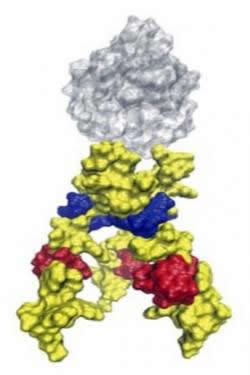
the structure of a key part of the HIV envelope protein, the gp41 membrane proximal external region (MPER), which previously eluded
detailed structural description.
The research will help focus HIV vaccine development efforts, which have tried for decades to slow the spread of a virus
that currently infects more than 33 million people and has killed 30 million more. The team reported the findings online in the Jan. 13
early edition of Proceedings of the National Academy of Sciences.
"One reason vaccine development is such a difficult problem is that HIV is exceptionally good at evading the immune
system," said Bruce Donald, an author and professor in Duke's computer science and biochemistry departments. "The virus has all
these devious strategies to hide from the immune system."
One of those strategies is a dramatic structural transformation that the virus undergoes when it fuses to a host cell. The envelope
protein complex is a structure that protrudes from HIV's membrane and carries out the infection of healthy host cells. Scientists have
long targeted this complex for vaccine development, specifically its three copies of a protein called gp41 and closely associated
partner protein gp120.
The authors said they think about a particular region of gp41, called MPER, as an Achilles' heel of vulnerability.
"The attractiveness of this region is that, number one, it is relatively conserved," said Leonard Spicer, senior author and a professor
of biochemistry and radiology. In a virus as genetically variable as HIV, a successful vaccine must act on a region that will be conserved,
or similar across subtypes of the virus.
"Second, this region has two particular sequences of amino acids that code for the binding of important broadly neutralizing
antibodies," said Spicer. The HIV envelope region near the virus membrane is the spot where some of the most effective antibodies found
in HIV patients bind and disable the virus.
When the virus fuses to a host cell, the HIV envelope protein transitions through at least three separate stages. Its
pre- and post-fusion states are stable and have been well studied, but the intermediate step -- when the protein actually makes
contact with the host cell -- is dynamic. The instability of this interaction has made it very difficult to visualize using
traditional structure determination techniques, such as x-ray crystallography and nuclear magnetic resonance (NMR) spectroscopy.
That's where Duke's interdisciplinary team stepped in, solving the structure using protein engineering, sophisticated NMR
and software specifically designed to run on limited data.
First author Patrick Reardon spent years engineering a protein that incorporated the HIV MPER, associated with a membrane
and behaved just like gp41 in the tricky intermediate step, but was stable enough to study. Reardon, then a PhD student under Spicer,
is now a Wiley postdoctoral fellow at the Environmental Molecular Sciences Laboratory, a scientific facility in the Department of
Energy's Pacific Northwest National Laboratory.
The result captured the shape of the symmetric, three-part MPER in its near-native state, but the protein needed to be more than
structurally accurate -- it had to bind the broadly neutralizing antibodies.
"One of the most important aspects of the project was ensuring that this construct interacted with the desirable antibodies, and indeed,
it did so strongly," Reardon said.
The team validated the initial structure using an independent method of data analysis developed by Donald's lab, which showed alternate
structures were not consistent with the data.
"The software took advantage of sparse data in a clever way that gave us confidence about the computed structure," Donald said. It used
advanced geometric algorithms to determine the structure of large, symmetric, or membrane-bound proteins -- varieties that are very
difficult to reconstruct from NMR data.
Donald's lab has been perfecting the method for a nearly decade, and Donald said its application in this paper represents a culmination
of that work, demonstrating how the two-pronged approach can illuminate the structure of complex protein systems.
The next steps of this research have already begun. In December, Duke received a grant of up to $2.9 million from the Bill & Melinda
Gates Foundation to fund the development of an HIV vaccine that will build on these findings.
In addition to Donald, Reardon and Spicer, the paper's authors include Harvey Sage, S. Moses Dennison, Jeffrey W. Martin, S. Munir Alam
and Barton F. Haynes, all from Duke.
Funding for this research came from National Institutes of Health grants GM-78031 and GM-65982.
Citation: "Structure of an HIV-1 Neutralizing Antibody Target. A Lipid Bound dp41 Envelope Membrane Proximal Region Trimer." Patrick F.
Rearden, Harvey Sage, et al. Proceedings of the National Academy of Sciences, Jan. 13, 2014. doi:101073/pnas1309842111
###
Contact:
Erin Weeks
(919) 681-8057
erin.weeks@duke.edu
Duke University
Source:
http://today.duke.edu/2014/01/hivvaccinefinal
For more HIV and AIDS News visit...
Positively Positive - Living with HIV/AIDS:
HIV/AIDS News
|